Oxidants in Daily Life and Common Health Conditions
Atherosclerosis
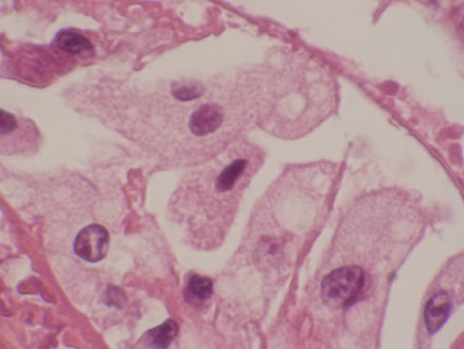
Lipids are transported in blood as lipoproteins, molecular complexes of various combinations of lipids with specific carrier proteins. Having polyunsaturated fatty acids as constituents, lipoproteins are a good target for free radical oxidation. Oxidized lipoproteins trapped in the arterial walls are an effective attractant of phagocytes, which gather around the lipoprotein deposits in an attempt to eliminate them. The phagocytes engorged with the lipids are now transformed into foam cells, so called because of their abundant lacy foamy cytoplasm of lipids (Fig. 3). Collections of these foam cells are detectable with eye over the inner surfaces of the arteries as orange yellow streaks, and represent early atherosclerosis.
The phagocytes thus activated also secret chemical substances that orchestrate and effect further inflammatory reactions, promoting the fatty streaks into full-blown atherosclerotic plaques over time. Atherosclerosis is now considered an inflammatory condition.
Free radicals� attacks on lipids are greatly facilitated in the presence of abundant reducing sugars, such as glucose, in the blood . Reducing sugar molecules attach themselves to lipids (glycation), altering the structural arrangement of the lipids in such ways that facilitate oxidative reactions, perhaps by giving free radicals a better access to their targets.
Free radicals� involvement in the pathogenesis of atherosclerosis is evident in the strong positive correlation found between smoking and atherosclerosis. Although we have antioxidants such as uric acid and indirect bilirubin in our blood, they are obviously not sufficiently effective in keeping smoking-related oxidative damages in check.
Indirect bilirubin is a powerful fat-soluble antioxidant that circulates in the blood attached to albumin. It is the most abundant antioxidant in mammalian tissue and responsible for most of the antioxidant activities in serum. When the oxidant concentration in the blood rises, an enzyme called Heme oxygenase-1 in the vascular lining cells (Endothelium) becomes activated and breaks down heme that comes from hemoglobin into bilirubin. This body�s response, however, is not swift enough and significant amounts of oxidative damage may have been incurred by the time the bilirubin level is up.
Further evidence implicating oxidants in atherosclerosis can be found in radiation induced -tissue injuries. In radiation therapies, oxidants are purposely induced within the body by applying energies of X-ray beams (see Ultraviolet and X-Rays) and these oxidants are the actual force utilized in the destruction of tumor cells.
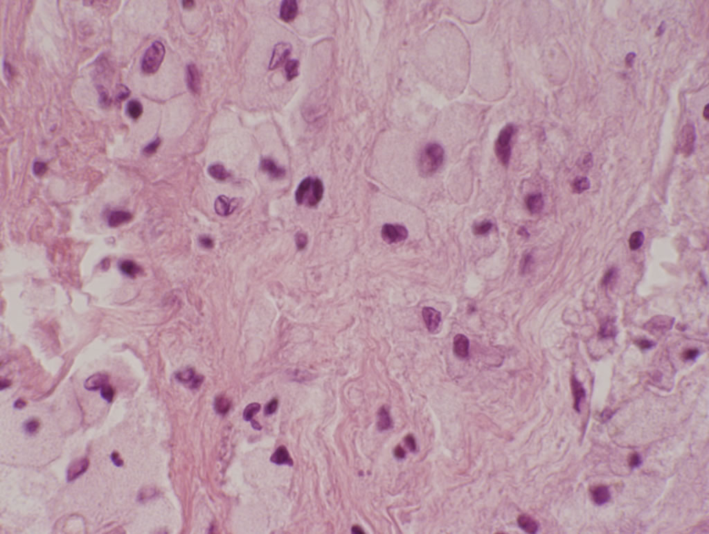
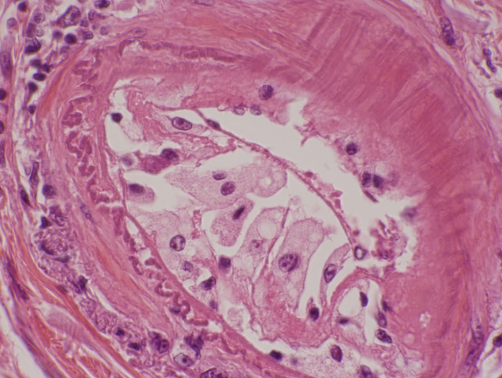
A biopsy or resected tissue taken from the area of the body that has been irradiated in the recent past often demonstrates foam cell collections in the walls of small arteries or arterioles (the smallest - sized arteries) that are similar to those seen in early - stage atheroscrelosis (Fig. 4). In fact, in a histological tissue examination looking for an evidence of radiation injuries, foam cell clusters in the arterial walls are one of the hallmarks of radiation effects. The foam cells are a telltale sign that the tissue has received radiation, either as an intended target or a bystander. In view of the fact that radiation works through oxidants, one can safely conclude that oxidants are causally related to the formation of foam cells. The artery thus affected eventually becomes narrowed from inflammation and fibrosis, just like in atherosclerosis, and can give rise to chronic radiation side effects, such as ulcer, bleeding or perforation of an viscus, due to poor blood circulation.
Copyright 2006 Kuma.us